Page content
The Cell Technologies Unit manages specialist equipment and laboratories for technologies involving the use and storage of mammalian cells and tissues.
These facilities include class II biosafety cabinets, CO2 incubators, temperature monitored and alarmed -80 freezers and vapour phase liquid nitrogen storage and flow cytometer.
The facility is capable of completing any aspect of tissue culture for external companies; this work is completed by the expert staff within the Cell Technologies Unit.
This facility has collaborated with several companies to test and evaluate compounds on human cell lines; as well as working on several projects staining and processing flow cytometry samples for local companies.
Facilities available through Cell Technologies Unit
Tissue culture and training
Mammalian Cell Propagation: A suite of tissue culture rooms is available for specialist propagation of a diverse range of mammalian cell lines (including stem cells) under normoxic and hypoxic conditions. This facility is capable of culturing cells for external companies; this can be used to test effects of compounds on cells in vitro. The facility can also further investigate the effects the compound is having on the cells to determine a mode of action. It is also possible to train users in the techniques commonly used in tissue culture.
Flow cytometry
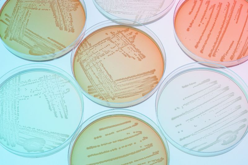
This facility has a two laser eight colour Beckman Coulter Gallios flow cytometer. The cytometer is capable of detecting compounds as small as 0.4mm. This level of detection allows the cytometer to investigate bacteria. Flow cytometry has many varied uses and these routinely include:
- Using fluorescent antibodies to identify and enumerate different cell types
- Immune function
- Investigation of protein levels within different cells types
- Apoptosis and necrosis analysis
- Cell cycle analysis
- Biomarker discovery
- Stem cell analysis
- Pharmaceutical analysis
- Plant cell biology
- Marine biology
Patch clamp
To record neuronal activity and neuron reconstruction. This facility is equipped with a specific confocal microscope to aid the analysis of neuronal activity in the slice (in vitro) which is recorded by the patch clamp; and a special cell filling system to label neurons, which are then analysed within our Bioimaging Facility.
Window chamber/skin flap model
Uses include in vivo imaging of vasculature, neurons and plaques in mouse models of cancer and Alzheimer's Disease (AD), using an in vivo window chamber system. Enables the visualization and quantification of various parameters of the circulation (plasma, erythrocytes and leukocytes) labelled with fluorescent dyes in real-time.
A diversity of cell types can be labelled, such as endothelium, grafted tissue (pre-labelled tumour cells) or specific cell markers (e.g. macrophages), enabling individual cell behaviour to be monitored in real time.The technique can be used to quantify physiological parameters of the vasculature such as flow, erythrocyte and leukocyte velocity and flux across vessel walls (useful in studies of drug kinetics in vivo).
We are experienced in studies involving tissue regeneration in the panniculus carnosus (pc) muscle as well as investigations of tumour vasculature in xentotransplanted tumours in this model. Other sites include the surface of the brain, where the interaction between vasculature and neurons can be examined in a mouse model of AD.
Cell and tissue storage
Mammalian cells and tissue are stored and maintained using a barcoded computerized system that adheres to the requirements of the Human Tissue Act (HTA). The facility has 24/7 monitored and alarmed -80 freezers and vapour phase liquid nitrogen storage. This provides a secure storage environment for samples.
This Unit also has access to the following standardised laboratory equipment:
- Thirty -80° freezer units in a temperature controlled environment storing up to 1.2 million samples
- Item tracker labelling and traceability software
- AAW WebReact Temperature Monitoring System
- Cell Radiation: Including Xray sources
- Countess Automated Cell Counter: Easy, accurate cell counting without the need for a hemocytometer
- Specialist Cell Imaging: Leica TCS SP5 Confocal Microscope for multiphoton imaging
- FLEXstation microphysiometer: For measuring transmembrane potentials and intracellular calcium signalling
Scientific Assistant

Dr Keith Thomas
Core Technologies Officer (Cell Technologies)