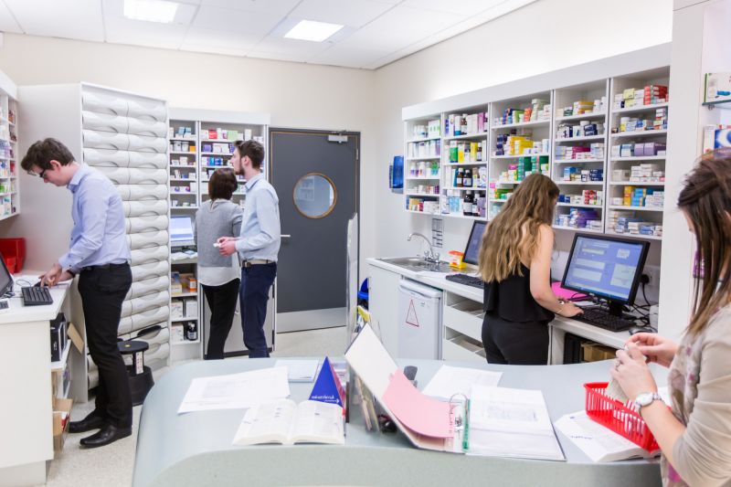
Page content
Retinal and Anterior segment OCT

We are currently involved in research using novel ophthalmic imaging including:
- Binocular OCT development in collaboration with Moorfields Eye Hospital.
- Confocal corneal imaging (e.g. in AK)
- We have investigated retinal profile and thickenss in ASD using retinal OCT scans.
- We are using both retinal and anterior segment OCT imaging to investigate retina in ASD, and to investigate the utility of OCT for biometric analysis and quantifying cataract magnitude.
In-vivo photoreceptor imaging
In-vivo photoreceptor imaging: we are investigating cone imaging using narrow angle Heidelberg Retinal Angiograph in the ageing eye and in ocular diseases such as glaucoma and diabetes.
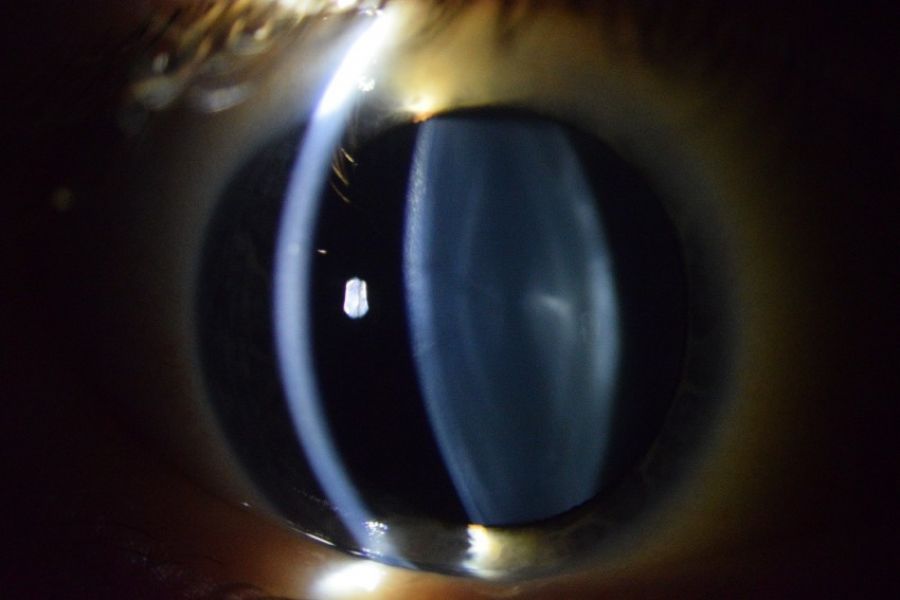
Slit lamp Photography
Our group have developed a bespoke slit-lamp based anterior segment imaging system and have employed this in a study investigating the profile of cataracts in Down syndrome.
We routinely capture high resolution fundus photography to investigate structure –function questions
Confocal Corneal imaging
Dr Mulholland employs Confocal corneal imaging in association with research at Moorfields Eye hospital