Page content
Northern Ireland Functional Brain Mapping Facility
Funded by Invest Northern Ireland and Ulster University, the functional brain mapping (FBM) facility at the ISRC is equipped with the latest whole head 306 channels Elekta Neuromag MEG TRIUX system. This is the only such facility on the island of Ireland.
MEG is a relatively modern non-invasive neurophysiological technique for measuring magnetic fields generated by neuronal activities inside the brain. Since the signal from the brain is much weaker than the ambient magnetic noise in the environment, the MEG facility is housed in a special magnetic shielded room.
The Northern Ireland FBM is part of a synergistic collaboration among the ISRC, School of Medicine (Personalised Medicine Centre), and Clinical Translational Research and Innovation Centre (C-TRIC).

- MEG provides a direct measure of electrical activity in the brain.
- MEG has a very high temporal resolution in the order of milliseconds (ms).
- MEG also has an excellent spatial resolution and is capable of localizing sources with an accuracy of millimetres (mm).
- It is a non-invasive technique which does not require any contrast injection.
- It is comfortable for subjects/patients. Unlike MRI, MEG does not produce any noise during scanning
In clinical applications, MEG is used for pre-surgical evaluation of brain tumour, arteriovenous malformation (AVM) and epileptic focus.
In research, MEG is known to produce better localized responses in the brain as a result of a better signal-to-noise ratio compared to EEG and a more straightforward biophysical formulation. It is widely used to study cognitive processes.
Although less explored but it can be used for the development of Brain-Computer Interfaces, and an analogue to EEG applications. In this case, it can be used essentially to inform EEG about effective biomarkers and learn better about brain dynamics using more active stimulation tasks.
Our Magnetoencephalography (MEG) laboratory is equipped with the latest whole head 306 channels Elekta Neuromag MEG TRIUX system. MEG is a modern non-invasive neurophysiological technique for measuring magnetic fields generated by neuronal activities inside the brain.
The MEG activity is acquired by an array of superconducting quantum interference devices (SQUIDs) placed close to the scalp which is capable of measuring magnetic fields on the femtotesla (fT) scale. Since the signal from the brain is too weak (~ 10-13 T) compared to the ambient magnetic noise in an urban environment (~ 10-7 T), MEG needs a special shielding from external magnetic signal. For this purpose, our MEG system is housed in a magnetic shielded room (MSR) for reducing noise from the surrounding environment.
Laboratory Facilities
We offer several techniques for brain stimulation and analysis that allow us to obtain better results and guarantee a good service for both research and clinical applications. Our MEG services can be booked using our NIFBM Booking System, which will soon be made available here.
Our lab is equipped with an MEG compatible BrainCap of 128 high-quality Ag/AgCl EEG electrodes from Brain Products GmbH. Electrodes are fixed to the cap and extremely flat which makes BrainCap very comfortable for the subject (e.g. avoiding excessive pressure onto the scalp, even in sleep studies during which the subject lies on the electrodes).
All EEG electrodes are buttoned directly into the cap (total height 3.5 mm) or can be attached to the skin with washers. This device facilitates concurrent MEG-EEG recording that allows an effective measurement of the brain activity.
Earphone for Auditory stimulation
Auditory stimulation is provided using an earphone, which is especially designated for MEG applications.
The stimuli are provided using stimulation programs that are installed in the STIM computer (STIM2) outside the MSR. The stimuli are applied with high precision having an extremely small delay of less than 20 milliseconds.
Visual stimulation
Visual stimulation is provided from the STIM computer through a Panasonic projector model PT-DS12KE (placed outside the MSR) projected onto a MEG compatible Elekta screen located inside the MSR. This allows a clear representation of the different scenarios of visual stimuli tasks, while keeping the image presentation delay shorter than 50 milliseconds.
Electrical stimulation
Electrical non-harmful stimulation is provided with small foam pads to stimulate instantaneously the median nerve on the Left/Right wrist. This stimulation will cause the thumbs to twitch and a trigger which is simultaneously recorded with the MEG signal.
Motor or response pads
We also have MEG compatible motor or response pads. Volunteers/patients will be asked to finger-tap the motor pad for performing Magnetic Evoked Field (MEF) tests. These pads can also be used as a response button for cognitive tasks.
We offer high quality neuro-imaging for both clinical and brain research purposes.
We adhere to highest standards of clinical practice. Following are the main highlight of our practice:
- MEG SQUID sensors are regularly checked for signal quality and are tuned and calibrated using phantom on a weekly basis. Also, a quick signal quality check is performed before every recording session.
- Before the commencement of recording, MEG staff provide a briefing about the MEG characteristics, requirements for the execution of mental tasks and what patients/subjects should do and avoid during the performance of a particular study task.
- Patients/volunteers will be thoroughly screened to check that they are suitable for the MEG recording (e.g. screening for possible metallic implant, braces, surgical aneurysm clips, etc., also for medical conditions that might disqualify them to perform certain mental tasks, while in the MEG scanner).
- After successful screening, the subject preparation is done on a special chair or MEG compatible bed. For concurrent MEG-EEG recording, subjects need to wear a 128 EEG channels BrainCap and will undergo the same preparation as for normal EEG recording. It might take up to 1 hour for EEG preparation. We may skip this part if the study only requires MEG recording as explained next.
- As part of the preparation for MEG recording, 4 or 5 head position indicator (HPI) coils will be attached to the subjects’ head using durapore tape. These coils allow to track head movement during the scanning. Then HPI coils and their whole head will be digitised using staylus pen in order to obtain a head shape for possible co-registration with their brain anatomical image. It might take approximately 15-30 minutes for MEG preparation and digitisation.
- Finally, the subjects will then be taken into the MSR and sit on a MEG chair or lay down on MEG bed and their head will be placed inside the MEG helmet as close as possible to the top of the helmet surface. They will be asked to keep still and be relaxed while performing the study tasks during the recording.
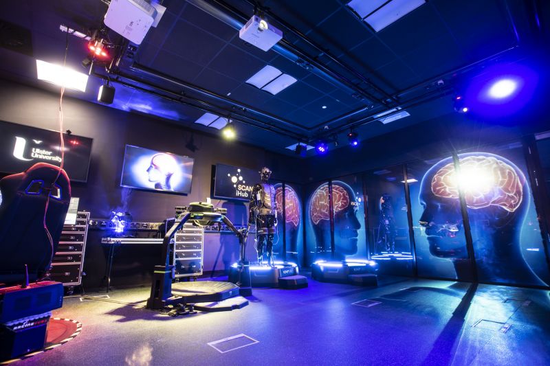
Spatial Computing and Neurotechnology Innovation Hub
The Spatial Computing and Neurotechnology Innovation hub (SCAN iHub) integrates our neurotechnology R&D with the latest development in human machine interaction and contains the latest technologies associated with these two computing areas enabling cutting edge research.It also enables new demonstrators for how this technology is utilised for training, large scale simulation, and immersive analytics.
SCAN iHub research focuses on developing capacity in “post-mobile” behaviours and modalities (e.g., text, voice, gesture, Augmented Reality/Virtual reality (AR/VR), and new contexts for computing (e.g., wearable and technology everywhere) with a unique focus on wearable electrophysiology and neurotechnology integration with the latest spatial computing technology.
The spatial computing and neurotechnology innovation hub (SCANi-hub) integrates our neurotechnology R&D with the latest development in human machine interaction and contains the latest technologies associated with these two computing areas enabling cutting edge research. It also enables new demonstrators for how this technology is utilised for training, largescale simulation, and immersive analytics.
SCAN i-hub houses multiple technologies for electrophysiological monitoring (multiple mobile wearable EEG headsets 1x64 channel and 2 x 32 channels EEG devices, a functional near infrared spectroscopy (fNIRS) brain imaging headset, ECG, HRV, pulse oximeter, GSR, and technologies for spatial computing including text, voice, gesture, augmented reality/virtual reality (AR/VR), an advanced car/flight simulator and vibrotactile stimulation suits, ultrasonic haptic interfaces and technologies that enable walking in a virtual environments (virtual treadmills) as well as a state-of-the-art Smartglass façade to adapt the room for various experimental situations and public engagement activities.
SCANi-hub research focuses on developing capacity in “post-mobile” behaviours and modalities (e.g., text, voice, gesture, Augmented Reality/Virtual reality (AR/VR), and new contexts for computing (e.g., wearable and technology everywhere) with a unique focus on wearable electrophysiology and neurotechnology integration with the latest spatial computing technology.
We were honored to welcome HRH Princess Anne to the Magee campus to officially open another new facility established at the ISRC, the Spatial Computing and Neurotechnology Innovation-Hub (SCANi-hub). The SCANi-hub builds on award-winning neurotechnology research at the ISRC and enables research in the next wave of human-computer and human-machine interaction for able-bodied and physically impaired people.
The facility sits alongside the Northern Ireland Functional Brain Mapping Facility (NIFBM) and houses multiple neurotechnologies for brain imaging, an advanced car/flight simulator, various new extended reality and spatial computing technologies, vibrotactile stimulation, ultrasonic haptic interfaces, technologies that enable walking in virtual environments (virtual treadmills) as well as a state-of-the-art Smartglass façade to adapt the room for various experimental situations and public engagement activities.
The SCANi-hub will equip the next generation of graduates and researchers with the skills and knowledge to merge bio-inspired computing and AI and SCAN technologies to address many research and industry-led challenges that help define how humans interact with technology in the future.
Northern Ireland - High Performance Computing Facility
We partnered with Queen’s University Belfast to establish the Northern Ireland High Performance Computing (NIHPC) Facility.
A grant of £2.1 million was awarded from the Department for Business, Energy and Industrial Strategy (BEIS) via the Engineering and Physical Research Council (EPSRC) and its national Tier-2 HPC initiative for the state-of-the-art computing facility. The Tier-2 HPC services are an interfacing layer of supercomputing provision between the highest capability national and international HPC services and local computing provision in individual research institutions.
Only one of seven such facilities in the UK, NI-HPC will enable researchers to conduct largescale data analytics, simulation and optimization of AI technologies to significantly enhance research productivity and quality, and to address some of society’s biggest challenges. An aim of the project is to develop the HPC software engineering knowledge and skills in Northern Ireland by providing hands-on technical training, coaching sessions and promoting the adoption of best practices.
We expect this to have a huge impact across a range of disciplines at both universities and industry sectors across Northern Ireland. The funding also enabled us to recruit Ulster’s first dedicated Research Software Engineer, Dr Jose Sanchez-Bornot, who is based at the ISRC.
Brain-Computer Interface (BCI) Laboratory
Our comprehensive BCI lab is equipped with:
- An electromagnetic field shielded room
- A state-of-art BCI experimental setup with 56 EEG channels, and 8 EMG channels (g.BSamp system)
- 4/8 EEG/EMG channel mobile unit (g.MOBIlab)
- A state-of-art 16-channel USB amplifier-based EEG/EMG/ECG/EOG mobile unit (g.USBamp)
- Multiple electrode systems: passive, active and dry electrodes
- A comprehensive bio-signal acquisition and processing system for measuring respiration, heart rate, galvanic skin response, skin temperature, oxygen saturation and eye-gaze
- A computerised smart wheelchair system for mobility control test
XR (Extended Reality) Health Hub
As the healthcare sector adopts and adapts to rapid technological advancement, XR is expected to be increasingly important with a broad range of use cases and applications. With advancements in XR technology and the subsequent emergence of new applications evaluating, personalising, and optimizing the user experience will be crucial to its widespread adoption.
Biometrics research has the potential to assess and enhance the user experience by leveraging physiological data to monitor emotional and cognitive responses to XR environments e.g., electroencephalography (EEG), electrooculography (EOG), electromyography (EMG) and photoplethysmography (PPG) can be used to measure brain activity, eye movements, muscle activity and heart rate. This data can be analysed in real-time to evaluate and adapt the XR experience and create more immersive and compelling content and interfaces tailored to the user's needs and preferences.
The XR (Extended Reality) Health Hub provides access to research, expertise, software development and demonstrations and equipment with a focus on health in state-of-the-art equipment facilities.